
Super-resolution microscopy by ROCS
| Background and Problem: Living cells are highly dynamic systems with cellular structures being often below the optical resolution limit. Super-resolution microscopes, usually based on fluorescence cell labeling, are usually too slow to resolve small, dynamic structures. We present a label-free microscopy technique, which can generate thousands of super-resolved, high contrast images at a frame rate of 100 Hertz and without any post-processing Approach: Our technique is based on oblique sample illumination with coherent light, an approach believed to be not applicable in life sciences because of too many interference artifacts. However, by circulating an incident laser beam by 360° during one image acquisition, relevant image information is amplified. By combining total internal reflection illumination with dark-field detection, structures as small as 150 nm become separable through local destructive interferences. The technique images local changes in refractive index through scattered laser light and is applied to living mouse macrophages and helical bacteria revealing unexpected dynamic processes. 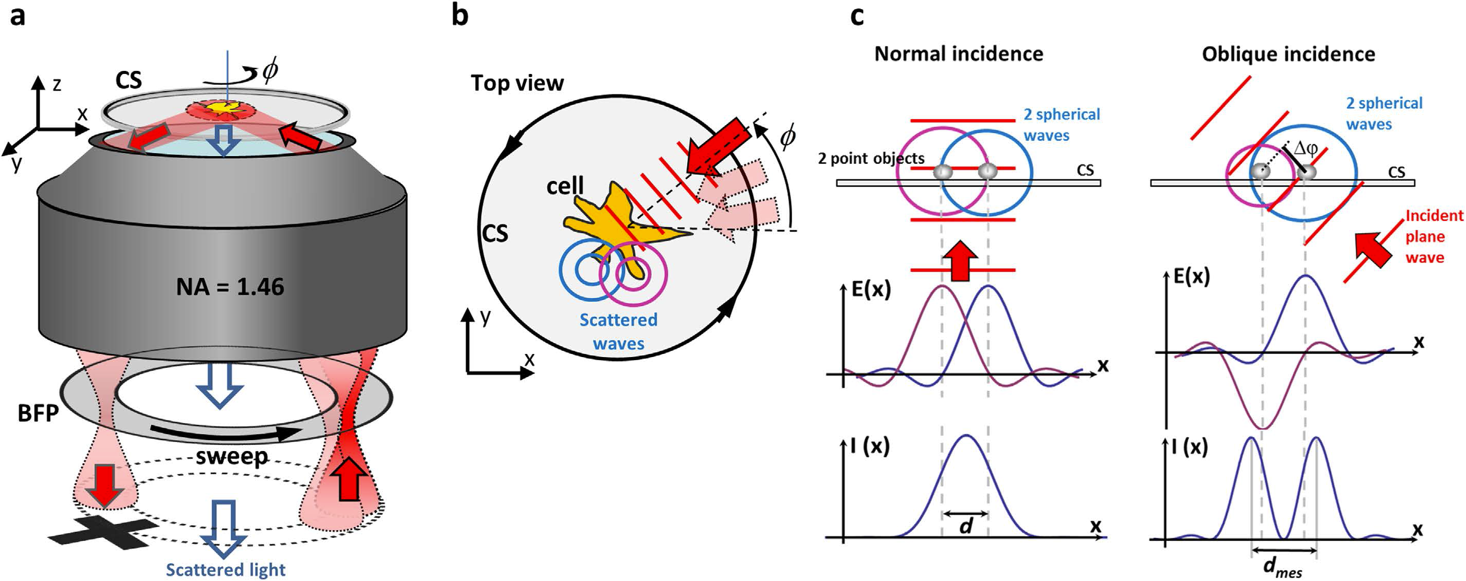 Experimental setup and operation principle. (a) A focused laser beam is scanned along a circle in the back focal plane (BFP) of the objective lens. The object on the coverslip (CS) is illuminated by an oblique plane wave, which is totally internal reflected (TIR-Mode). The scattered light is transferred to the camera, whereas the reflected beam is blocked to enable dark field imaging. (a,b) Within the integration time of the camera, the laser sweeps a φ = 0…2π angle. (b) Each plane wave, indicated by the red wavefronts, is multiply scattered at the object (the cell), indicated by two spherical waves (in blue and purple). For each angle φ, a (radially) coherent image with many artefacts is generated. A superposition of many different coherent images within the integration time of the camera generates a (tangentially) incoherent image with hardly any artefacts. (c) For normal incidence, two scatterers in a short distance d < λ/2 excite two spherical waves, whose electric fields E(x) are in phase at the camera. The two points cannot be resolved by the resulting intensity I(x) at the camera. For oblique incidence, the two scatterers excite spherical wavefronts with a phase delay Δϕ = π. Two maxima in a distance dmes can be resolved in I(x). |
Figures
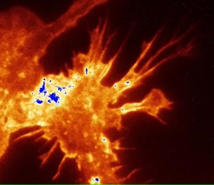
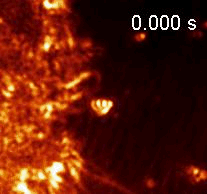 Fast cytoskeleton reorganization inside a macrophage upon interaction with 350nm beads (imaged at 63Hz with 150nm resolution).
|
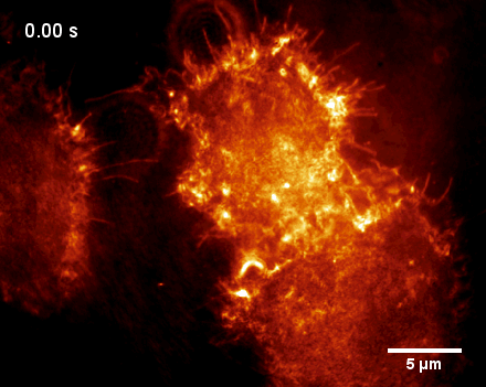
TIR-ROCS time series of J774 macrophages, exhibiting rich cytoskeleton dynamics. The cells use their dynamic filopodia to sense their surroundings as well as cell-cell-contacts. The dynamics can be imaged over a long period of time with consistently high image contrast.
| |
| ||
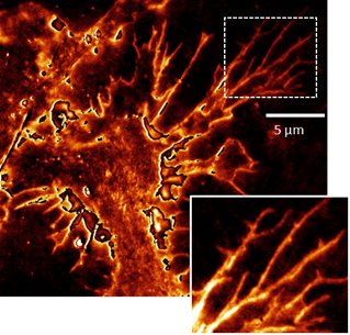
| Static TIR-ROCS image of a J774 cell (left image) with adherent filopodia that can be imaged at superior resolution and image contrast compared to the TIRF image (lifeAct-GFP actin staining, right image). | ||
Publications
- Ruh D, Mutschler J, Michelbach M, Rohrbach A
Superior contrast and resolution by image formation in rotating coherent scattering (ROCS) microscopy
2018 Optica, Band: 5, Nummer: 11, Seiten: 1371 - 1381 - Koch M D, Rohrbach A
Label-free imaging and bending analysis of microtubules by ROCS microscopy and optical trapping
2018 Biophys J, Band: 114, Nummer: 1, Seiten: 168 - 177 - Jünger F, Rohrbach A
Strong cytoskeleton activity on millisecond timescales during particle binding and uptake revealed by ROCS microscopy
2018 Cytoskeleton, Band: 75, Seiten: 410-424 - Jünger F, Olshausen P, Rohrbach A
Fast, label-free super-resolution live-cell imaging using rotating coherent scattering (ROCS) microscopy
2016 Sci Rep-uk, Band: 6, Seite: 30393 - Olshausen P, Rohrbach A
Coherent total internal reflection dark-field microscopy: label-free imaging beyond the diffraction limit
2013 Opt Lett, Band: 38, Nummer: 20, Seiten: 4066 - 4069